


Nanobacteria
What is Nanobacteria?
Nanobacteria might be possibly the oldest living buffering system within a body. There is much controversy surrounding nanobacteria. Is it alive? Is it mineral? Is it a cross between minerals and bacteria? Some have chosen to call them nanobacteria others prefer the term nanoparticles.
Nanobacteria or Nanoparticles
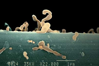
Some new research points to electron microscope size bacteria known as nanobacteria to be a living buffering system of the body. Nanobacteria or nanoparticles exist in two states. This first is a slime like state. These tiny electron microscope size bacteria spring into action when the pH of the body turns slightly acidic. They begin immediately to capture acid minerals and form these acid minerals into stones. This further assists the body to maintain a delicate balance of neutral, neither acid nor alkaline. These stones, in theory, line the blood vessels and are commonly known as plaque. These mineral stones, in theory, form kidney stones. In theory these mineral stones create inflammation in polycystic kidney cells. If we can somehow manage this inflammation, it is thought in theory, that we may be able to stop the spiraling downward progression of kidney decline in polycystic kidney disease.Nanoparticles' effect on our bodies, can be changed through alkalinity, through diet and lifestyle.
One theory on nanobacteria, is that they might possibly be among the oldest, the very first organisms to have ever exist. They have found nanobacteria on carbon dated age old rocks. Nano particles appear to be a cross between mineral and bacteria. Nanobacteria could be the only living pH regulators that exist in this world today. They go easily from their worm like or slime form into their apatite mineral rock form. Nano present as a slime in order to propel themselves along. If the host body, i.e. our body, becomes too acidic, nanobacteria jump into action and sacrifice themselves changing into rock like crystals that will bind the body's acids. It is a great protector that helps us to survive; nano is a great help to our bodies; it tries very hard to get us to survive; nano help us to remain neutral alkaline. That is the purpose of nanobacteria.
They do form kidney stones if the body becomes too acidic. They do form blood vessel plaque if the body is too acidic. This stone formation is nano's way of capturing the acidic minerals. Nanobacteria senses that the body's pH is becoming dangerously acidic, so it springs into action, changing its form from a worm like micro organism propelling its self in a slime like state changing to a rock filled with acidic minerals. This rock binds all available acids that can possibly be bound. The body stays alive. The body stays neutral. The body survives. We can alter this by helping our own bodies to remain alkaline. Generally with alkalinity, there will be no kidney stone formation. Generally with alkalinity there is no blood vessel plaque.
" Nanobacteria are unconventional agents 100-fold smaller than common bacteria that can replicate apatite-forming units. Nanobacteria are powerful mediators of biogenic apatite nucleation (crystal form of calcium phosphate) and crystal growth under conditions simulating blood and urine. Apatite is found in the central nidus of most kidney stones and in mineral plaques (Randall's plaques) in renal papilla. The direct injection of nanobacteria into rat kidneys resulted in stone formation in the nanobacteria-injected kidney during one month follow-up, but not in the control kidney injected with vehicle. After intravenous administration in rats and rabbits, nanobacteria are rapidly excreted from the blood into the urine, as a major elimination route, and damage renal collecting tubuli. Nanobacteria are cytotoxic to fibroblasts in vitro. Human kidney cyst fluids contain nanobacteria. Nanobacteria thus appear to be potential provocateurs and initiators of kidney stones, tubular damage, and kidney cyst formation. It is hypothesized that nanobacteria are the initial nidi on which kidney stone is built up, at a rate dependent on the supersaturation status of the urine. Those individuals having both nanobacteria and diminished defences against stone formation (i.e. genetic factors, diet and drinking habits) could be at high risk. Kidney cyst formation is hypothesized to involve nanobacteria-induced tubular damage and defective tissue regeneration yielding cyst formation, the extent of which is dependent on genetic vulnerability."
Nanobacteria or Nanoparticles?
Int J Nanomedicine. 2012;7:339-50. doi: 10.2147/IJN.S28069. Epub 2012 Jan 19.
Kutikhin AG, Brusina EB, Yuzhalin AE.
Source
Department of Epidemiology, Kemerovo State Medical Academy, Kemerovo, Russian Federation. antonkutikhin@gmail.com
Abstract
Calcifying nanoparticles (CNPs) (nanobacteria, nanobacteria-like particles, nanobes) were discovered over 25 years ago; nevertheless, their nature is still obscure. To date, nobody has been successful in credibly determining whether they are the smallest self-replicating life form on Earth, or whether they represent mineralo-protein complexes without any relation to living organisms. Proponents of both theories have a number of arguments in favor of the validity of their hypotheses. However, after epistemological analysis carried out in this review, all arguments used by proponents of the theory about the physicochemical model of CNP formation may be refuted on the basis of the performed investigations, and therefore published data suggest a biological nature of CNPs. The only obstacle to establish CNPs as living organisms is the absence of a fairly accurately sequenced genome at the present time. Moreover, it is clear that CNPs play an important role in etiopathogenesis of many diseases, and this association is independent from their nature. Consequently, emergence of CNPs in an organism is a pathological, not a physiological, process. The classification and new directions of further investigations devoted to the role of CNPs in biology and medicine are proposed.
PKD Stones and Nanobacteria
Antimicrob Agents Chemother. 2002 Jul;46(7):2077-86.
Cíftçíoglu N, Miller-Hjelle MA, Hjelle JT, Kajander EO.
Source
Department of Biochemistry, University of Kuopio, FIN-70211, Kuopio, Finland.
Abstract
Compounds from 16 classes of antimicrobial drugs were tested for their abilities to inhibit the in vitro multiplication of nanobacteria (NB), a newly discovered infectious agent found in human kidney stones and kidney cyst fluids from patients with polycystic kidney disease (PKD). Because NB form surface calcifications at physiologic levels of calcium and phosphate, they have been hypothesized to mediate the formation of tissue calcifications. We describe a modified microdilution inhibitory test that accommodates the unique growth conditions and long multiplication times of NB. This modified microdilution method included inoculation of 96-well plates and determination of inhibition by periodic measurement of the absorbance for 14 days in cell culture medium under cell culture conditions. Bactericidal or bacteriostatic drug effects were distinguished by subsequent subculture in drug-free media and monitoring for increasing absorbance. NB isolated from fetal bovine serum (FBS) were inhibited by tetracycline HCl, nitrofurantoin, trimethoprim, trimethoprim-sulfamethoxazole, and ampicillin at levels achievable in serum and urine; all drugs except ampicillin were cidal. Tetracycline also inhibited multiplication of isolates of NB from human kidney stones and kidney cyst fluids from patients with PKD. The other antibiotics tested against FBS-derived NB either had no effect or exhibited an inhibitory concentration above clinically achievable levels; the aminoglycosides and vancomycin were bacteriostatic. Antibiotic-induced morphological changes to NB were observed by electron microscopy. Bisphosphonates, aminocaproic acid, potassium citrate-citric acid solutions, and 5-fluorouracil also inhibited the multiplication of NB in a cidal manner. Insights into the nature of NB, the action(s) of these drugs, and the role of NB in calcifying diseases may be gained by exploiting this in vitro inhibition test system.
Nanobacteria: Is it alive?